When a tumor, aneurysm or other abnormality develops in the base of your skull, it can impact your ability to see, hear, talk, move and think. We specialize in treatments, like minimally invasive surgery, including using endoscopes, to remove tumors or other growths to get you back to living the quality life you deserve.
Expert surgery for your peace of mind
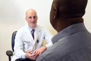
Learning that you need surgery can bring a wave of emotions. But we’re here to help you navigate these new waters with proper planning and a skilled, experienced team by your side.
For more than 30 years we have safely removed tumors from along the skull base while minimizing the impact on brain tissue. Your care team will work closely together, sharing your health history, current condition, and treatment plan to ensure comprehensive coverage of all your healthcare needs.
Depending on the location of your tumor or aneurysm, your team may include professionals from the following specialties:
Find a doctor near me
Conditions
Your care team may recommend skull base surgery to treat tumors and other abnormalities along the skull base, ears and around the eyes, including:
Acoustic neuromas (vestibular schwannoma)
Acute facial nerve paralysis
Adenoid cystic carcinoma
Aneurysmal bone cysts
Basilar invagination
Brain aneurysm
Carotid body tumors
Cavernous hemangiomas
Cavernous malformations
Cerebrospinal fluid (CSF) leaks through the nose or ear
Cholesterol granulomas
Chondrosarcoma
Chordoma of the clivus
Chordomas
Craniopharyngioma
Dermoid cysts
Dural arteriovenous (AV) fistulas
En plaque sphenoid wing meningiomas
Encephaloceles
Endolymphatic sac tumors
Epidermoid cysts
Esthesioneuroblastoma
Facial nerve schwannomas
Fibrous dysplasia
Glomus jugulare tumors and other paragangliomas
Glossopharyngeal and vagal schwannomas
Hemangioblastomas
Hemifacial spasm
Inner ear tumors
Intraosseous hemangiomas
Juvenile nasal angiofibromas
Meningiomas
Meningoceles
Neurofibromas
Orbital tumors
Osteomas
Pituitary tumors
Rathkes cleft cysts
Schwannomas
Semicircular canal dehiscence
Sinonasal cancer
Solitary fibrous tumors
Temporal bone tumors including malignancy
Trigeminal nerve schwannomas
Testing
Skull base surgery requires careful planning and diagnostic tests to get a clear picture of your condition and plan a safe and effective procedure. Tests may include:
- MRI/MRA scans take detailed pictures of your brain and blood vessels, helping us create the best surgical plan for you.
- CT or CTA scans provide clear images of your skull's bones and blood vessels, helping us understand the structure of your skull base.
- Audiograms are tests that check how well you can hear.
- Optical coherence tomography (OCT) is a non-invasive imaging test that evaluates your eye health.
- Cerebral angiography gives a detailed view of the blood vessels in your brain.
- Pituitary hormone testing checks how well your pituitary gland functions, which is important to identify any hormonal issues prior to surgery.
Treatments
Skull base surgery is usually just one part of a larger treatment plan that may include medication, "watchful waiting" with periodic MRI imaging, Gamma Knife surgery and radiation therapy.
Types of skull base surgery
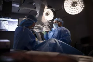
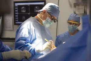
When performing skull base surgery, we prioritize minimizing your potential pain, risks, and recovery time. That's why our surgeons specialize in minimally invasive and endoscopic procedures. We can make surgical incisions roughly the size of a marble to remove tumors while sparing healthy tissue. Our tools include:
- Cerebral catheters, coils, stents, glues and flow diverters
- CO2 Laser
- Computer-guided surgical navigation systems
- Gamma Knife surgery
- High-definition endoscopes
- Surgical approaches that minimize brain movement
- Ultrasonic aspirator (CUSA)
We focus on helping people recover safely and comfortably after brain surgery. Our surgical team has a lot of experience with the most challenging and complex conditions. If your tumor needs traditional or "open" surgery, you can trust that you will be in the hands of top neurosurgeons.
FAQs
What are the risks of skull base surgery?
The risks of skull base surgery depend on the extent and location of the procedure. Common risks include infection, cerebrospinal fluid leakage, cranial nerve injury, postoperative bleeding, and complications from general anesthesia. Additional risks vary based on the tumor's size and location, the operation's duration, and your overall health. Fortunately, severe complications such as major strokes, pulmonary embolism, heart attack, paralysis, coma, or death are very rare.
How long will I be in the hospital after skull base surgery?
The length of your hospital stay will depend on the type of surgery you have. Endoscopic endonasal surgery, a minimally invasive technique using an endoscope inserted through the nostrils to reach the surgical site without external incisions, usually requires a 2-day stay in the hospital. However, this stay may be longer if there are concerns about postoperative cerebrospinal fluid leakage.
If you have a craniotomy with open microsurgery, you will generally go home after 2-3 days.
Will I need additional treatment after skull base surgery?
If a non-cancerous tumor (benign tumor) is completely removed, no other treatment is needed except for regular MRI scans to check that it hasn't come back. If the whole benign tumor can't be removed, the leftover part is usually treated with a special type of surgery called Gamma Knife radiosurgery. Sometimes, we may just watch the remaining tumor and only treat it if it starts to grow again.
Cancerous tumors at the base of the skull usually need radiation after surgery. This radiation can be done all in one day (radiosurgery) or spread out over several weeks (radiation therapy). Sometimes, chemotherapy or immunotherapy is also used, depending on the type of tumor.
Is acoustic neuroma cancer?
An acoustic neuroma (also known as vestibular schwannoma) is rarely a cancer. They typically do not spread to other body parts (metastasize), spread throughout the cerebrospinal fluid, or invade the brain.
Will my acoustic neuroma become cancer?
Acoustic neuromas turning into cancer is an extremely rare event.
How do you know that my tumor is an acoustic neuroma just by looking at the MRI?
Acoustic neuromas have a very typical appearance on an MRI scan and can usually be diagnosed without a tissue sample. The tumor location, shape, pattern on an MRI and widening of the internal auditory canal, all help doctors identify an acoustic neuroma on an MRI scan. On rare occasions, a facial nerve schwannoma will mimic the MRI appearance of an acoustic neuroma.
Will an acoustic neuroma cause me to go deaf?
There is a risk of losing hearing in your ear with an acoustic neuroma regardless of the treatment path chosen. The exact risk depends on many factors including hearing quality at diagnosis, the tumor’s growth rate, the tumor size, and the treatment given. There is also a risk of hearing loss over time without treatment if you undergo watchful waiting with periodic MRI imaging.
Will surgery for my acoustic neuroma help my tinnitus?
Surgery for an acoustic neuroma is not a realistic treatment for tinnitus. Some people may report that their tinnitus changed or decreased after treatment, but this is not an expected outcome of either surgery or Gamma Knife radiosurgery. The effect of treatment on your tinnitus is not something that can be expected or predicted.
What is the orbit?
The orbit is also known as the eye socket and includes all the bones that house the eye as well as the eye muscles, blood vessels, nerves, and other soft tissues.
What does an orbital surgeon do?
An orbital surgeon is an ophthalmologist (eye doctor) who completed additional training in a wide range of conditions affecting the areas that surround the eye, including the orbit or eye socket. They often partner with colleagues in neurosurgery and head and neck surgery to provide comprehensive care of your skull base.
What can I expect during endoscopic pituitary surgery?
Endoscopic Endonasal Resection (EER) is a minimally invasive surgery that uses an endoscope, a small camera, inserted through your nose to reach the skull base. Your surgeon will maneuver instruments to this area to open a small hole in the bone where the pituitary gland sits, in a depression called the Sella. This approach allows us to remove tumors of the pituitary gland and surrounding areas. EER treats various types of tumors, including pituitary adenomas and meningiomas.
Our locations
From regular office visits to inpatient stays, find the healthcare you need and deserve close to home.
Our doctors + care team
Meet the doctors and care team devoted to supporting you along your path to better health.