For more information about our services or to schedule a tour, please call 617.636.5613 or email Corinna.Beale@tufts.edu.
About
Wound healing
Ira Herman
Dr. Herman’s lab focuses on understanding the molecular mechanisms that control cellular growth and differentiation during the development or disease. Using the vasculature as a model system for in vitro and in vivo studies, the group implements morphologic, biochemical and molecular genetic approaches. Experimental results have yielded important new insights into the molecular and cellular regulators that control cell migration and division, angiogenesis and hypertension or wound healing. Studies performed at the SIRL focus on factors that modulate angiogenesis to regulate wound healing.
Mark Iafrati
Dr. Mark Iafrati is the chief of vascular surgery and the Director of the Center for Vascular, Would Healing and Hyperbaric Medicine at Tufts Medical Center. He is studying the mechanisms of wound healing and molecules that can accelerate the process in patients with diabetes.
Cardiology
Navin Kapur
As an Interventional Cardiologist and Advanced Heart Failure/Cardiac Transplant Physician, Dr. Kapur’s laboratory at the MCRI and SIRL focuses on preclinical models of left heart failure, myocardial infarction, ischemia-reperfusion injury and pulmonary hypertension. Unique aspects of the research program include biventricular pressure-volume loop analysis and 3D echocardiography in mice and large animals. According to the literature, Tufts SIRL holds the longest-standing ischemia model in pigs. Learn more about MRI 3D/STE.
Dental
Pamela Yellick
Dr. Yellick’s research on craniofacial and dental tissue development and regeneration research uses two approaches. The principles of tissue engineering, employing post-natal epithelial and mesenchymal dental stem cells, seeded onto biodegradable scaffolds and implanted in an in vivo model, to regenerate organized tooth crowns, periodontal tissues and whole teeth.
The laboratory also exploits the developmental model of the zebrafish, using a mutagenesis approach combined with an in vivo mineralized tissue stain, to identify craniofacial and primary and replacement tooth mutants. The synergistic effects of these two approaches provide a unique opportunity to identify genes regulating craniofacial and replacement tooth formation, and to test them in clinically relevant model systems.
Driss Zoukuri
Osseointegration of silk-coated implants
Replacement of missing teeth in dental clinics has always been a challenge in the dental field. Dental implants became the ultimate gold standard solution for permanently replacing missing teeth. For the implants to stay functional and replace dentition efficiently, osseointegration (defined as a union between bone and titanium surface without having a soft tissue barrier or fibrous encapsulation) must occur.
Improving the surface of titanium implants has been the quest of many research fields, and one approach would be coating implants with RGD peptide-decorated silk, which will attract fibroblasts and thus improve bone adhesion to implant surfaces.
Dr. Zoukhri’s studies are testing whether coating titanium implants with silk, would improve osseointegration of dental implants. Finding new and innovative biocompatible materials for implants has a broad impact in many fields of surgery. The SIRL facility provides an optimum atmosphere for the support of these in vivo studies.
Optimal duration of triple antibiotic application on teeth
Clinically, the presence of pulp inflammation and infection have been established to be determinants for the failure of pulpotomy. A combination of various antibiotics effectively minimizes the inflammatory responses of pulpal tissues used in treating pulp lesions. Both in vitro and in situ studies have shown the effectiveness of the triple antibiotics in disinfecting root dentin (middle layer of the tooth).
If triple antibiotics are used correctly for the right amount of time, repair of the damaged tissue can be expected. To date, there is no guideline for triple antibiotic application duration; however, if a shorter duration of triple antibiotic application is shown to be effective in disinfecting the tooth, this could prevent the development of drug resistance.
These studies aim to investigate the optimal duration of topical triple antibiotics application in teeth. This research is conducted in the SIRL facility. The SIRL team has helped in the development and execution of this surgical model of tooth infection to examine pulp response measured by the level of inflammatory cell response, hard tissue formation, tissue necrosis and bacterial infiltration, coupled with radiographic examination of periodontal ligament thickness.
Women's health
Michael House
Preterm birth affects over 12% of pregnancies in the U.S. and leads to significant newborn morbidity and mortality. In some cases, preterm birth is caused by early cervical shortening and dilation. Dr. House and his team are developing injectable biomaterials to prevent cervical shortening, which should prevent preterm birth. These novel biomaterials are being tested in the SIRL in an animal model of pregnancy.
Ophthalmology
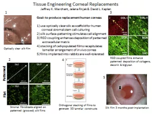
Jeffrey Marchant, PhD
Dr. Marchant's lab's goal is to develop a replacement human cornea. To this end, he uses silk-based scaffolds seeded with human corneal stromal stem cells. The scaffolds possess surface morphological characteristics that promote cell alignment and ordered deposition of extracellular matrix molecules. Orthogonal layers of seeded silk films mimic the endogenous corneal organization and are transparent. Slow silk degradation of the implanted constructs over time will lead to the complete replacement of the construct with native tissues.